The Eye That Winked Without Permission
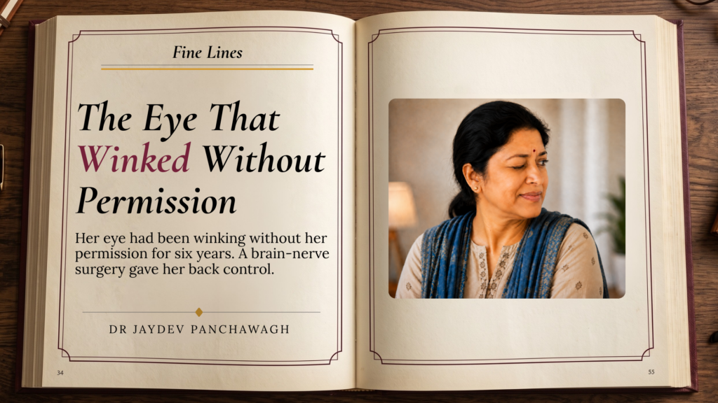
In This Essay A Twitch Called Hemifacial Spasm The Surgery Behind It: Microvascular Decompression Recovery After Microvascular Decompression We spend our lives learning to read faces without being taught. A raised eyebrow. A flicker at the corner of the mouth. Long before Darwin wrote The Expression of the Emotions in Man and Animals and catalogued the muscles that betray us, humans already knew that the face speaks a language older than words. It is why an actor can hold a stage with nothing but stillness, and why a single twitch, in the wrong moment, can undo an entire sentence. Most of us assume the face obeys us. It does — mostly. But there is a small, stubborn category of movement the face makes entirely on its own. A wink that isn’t a wink. An eye that closes when nothing has asked it to. For most people this passes in seconds, a minor mutiny, forgotten by lunch. For a woman I will call Meera, it did not pass. It had been happening for six years. A Twitch Called Hemifacial Spasm A flicker at the corner of the eye, and the years it took to be taken seriously Meera arrived at my clinic with her husband, a folder of prescriptions, and a habit she had developed without noticing — angling her face slightly away from whoever she was speaking to, so the left side, the side that moved, stayed out of view. It had started small. A flutter near the eye, the kind everyone gets after too little sleep or too much coffee. Hers did not go away. Over months, it spread — from the eyelid down to the cheek, then to the corner of the mouth, until one side of her face was, in her own words, “doing its own thing.” This is Hemifacial Spasm — involuntary, irregular contractions on one side of the face, caused not by nerves but by something touching them. Before we reached that word, Meera had been told it was stress. Then eye strain. Then, gently, that it might be anxiety, and had she considered speaking to someone. None of this is unusual. Hemifacial Spasm is often misdiagnosed for years, mistaken for a tic, a psychological symptom, or the ordinary wear of a stressful life, because on the surface, a twitch looks like nothing at all. It takes a particular kind of attention to notice that the twitch is not behaving like an ordinary one — that it never announces itself with fatigue or resolves with rest, that it has a rhythm too regular to be voluntary. Darwin argued that facial expression is not learned, but wired into us before birth, shared across cultures and species. He never imagined that in a few people, that same wiring could misfire — not from emotion, but from a blood vessel resting somewhere it shouldn’t. The Surgery Behind It: Microvascular Decompression Where the trouble actually begins — not in the face, but deep behind it The facial nerve does not begin at the face. It begins at the brainstem, deep at the base of the skull, where it exits as a thin, pale thread no thicker than wet string. In Hemifacial Spasm, a blood vessel — most often a small branch of the arteries supplying the brainstem — comes to rest directly against this nerve, at the exact point where it leaves the brain. This vessel does not sit still. It pulses, once with every heartbeat, thousands of times a day, brushing against the nerve with each beat. Oliver Sacks would have recognised the shape of this story immediately — a case where the villain is not a disease in the usual sense, but a heartbeat in the wrong place. His book The Man Who Mistook His Wife for a Hat is full of such cases. Each one shows a nervous system misfiring in some small, specific way. Each patient is stranger, and more human, than any textbook entry could make them. Hemifacial Spasm belongs on that shelf. Over months and years, the repeated, rhythmic contact of vessel against nerve irritates the nerve fibres until they begin to misfire, sending signals to the facial muscles that the brain never intended. The twitch a person sees in the mirror is, quite literally, an echo of their own pulse. The operation to correct this — Microvascular Decompression — does one thing, and it does it precisely. Under an operating microscope, magnified many times over, we find the vessel and free it from the nerve. We lift it away from the point of contact and hold it there, permanently, with a small piece of Teflon — inert, soft, and biologically unreactive — placed between vessel and nerve like a doorstop that never rusts. With continuous nerve monitoring running throughout the operation, we can watch the nerve’s electrical behaviour change in real time, confirming the exact moment the irritation stops. The compression that had been running quietly beneath her skin for six years is separated from the nerve in minutes. The spasm has one cause. Remove the cause, and the spasm has nothing left to answer to. For most patients, the change is not gradual. It is immediate — on the table, or within the following days. The muscles that had been contracting to their own private rhythm simply stop. Recovery After Microvascular Decompression What it means to look straight into a camera and recognise every part of what looks back Meera kept one photograph from before the surgery, tucked at the back of a drawer. It was from a wedding, taken three years into the spasm. She is standing at the edge of the frame, half turned, her chin dropped just enough to catch the light differently. Nobody else in the photograph is doing this. She remembers exactly why she did. For six years, every photograph was a small negotiation: which angle, how much light, whether to laugh with her mouth closed. Her husband had made calls, and visited other doctors, before they ever came
The Body Speaks First
I have spent much of my working life looking directly at the brain. Under the microscope it does not look like a mind. It is a tissue — pale, glistening, threaded with vessels and neurons finer than hair. Somewhere in that tissue lives everything a person is: their memory, their grief, their capacity for joy, their creativity, their essence!. The person who walks into my clinic and the organ that I look at later are the same. One is integrated into the other, yet somehow, sometimes feel different. This gap, between the felt self and the physical organ, is where yoga, exercise, and breath do their quiet work. It is also where a great deal of modern brain health research now lives. What Neurosurgery Teaches About the Mind The brain is responsive. Neurosurgery teaches humility about this gap. I have operated on brains that looked structurally identical under the microscope and yet belonged to people living entirely different inner lives. Anatomy alone does not explain temperament, resilience, or despair. Somewhere between the wiring and the person, physiology and psychology cross paths continuously, invisibly, all day. This is not new knowledge. In fact, it is as old as the ancient scriptures, and perhaps older. Long before imaging could show a hippocampus changing shape or a vagus nerve firing, the Yogis had already worked out, through daily observation and discipline, that the body was never separate from the mind. Modern neuroscience has mostly caught up to confirm what they intuited centuries ago. The Two-Way Street: Psychosomatic and Somatopsychic The mind moves the body. The body moves the mind back. Most of our actions-conscious and subconscious are psychosomatic: the mind acts and controls the body. Anxiety tightens the chest. Grief exhausts the limbs. Chronic stress raises cortisol (the stress hormone) and, over years, changes how the brain itself is built. This direction is well accepted, well studied, rarely questioned. The reverse direction, somatopsychic, is spoken about less, though it is equally real. The body also acts on the mind. A stiff, sedentary body tends to carry a duller mood. A body that moves regularly — through yoga, walking, swimming, any sustained activity — tends to carry a steadier one. Body and mind are not two systems talking to each other. They are one system, described in two languages. The nervous system does not read this as metaphor. Movement changes blood flow to the brain, alters circulating hormone levels, and adjusts the tone of the vagus nerve — the long, wandering nerve that links gut, heart, lungs, and brain in constant two-way conversation. Yoga, Exercise, and the Ageing Brain Time asks a question of every brain. Movement is the honest answer. Every year, research adds to a fairly consistent finding: people who stay physically active in midlife and beyond tend to show better preservation of brain volume, particularly in the hippocampus, the structure most involved in memory. Regular aerobic activity is associated with higher circulating levels of a protein called brain-derived neurotrophic factor, or BDNF, which supports the survival and growth of neurons. Regular practice is also linked to neuroplasticity — the brain’s ability to keep forming new connections, not only in childhood but across life. Several large population studies suggest that sustained physical activity, including yoga-based movement, is associated with a lower risk of cognitive decline and dementia in later life. I want to be careful here: Association is not proof of prevention. No form of exercise, yoga included, guarantees protection against dementia. What the evidence honestly supports is a lower risk, and better quality of life. Breath as the Body’s Oldest Medicine Of all the involuntary things the body does, breath alone will let you hold the wheel. Breathing is unusual among bodily functions. The heart cannot be consciously slowed by an act of will. Digestion cannot be willed faster. But breath sits right at the border of voluntary and involuntary, and yogic tradition discovered this border early and built an entire discipline around it. Slow, controlled breathing activates the vagus nerve and shifts the body toward its parasympathetic state — the rest-and-repair mode — lowering heart rate and easing the release of stress hormones. This is measurable now, in heart rate variability studies and in functional imaging. Pranayama is not mysticism dressed up in the language of science. It is a very old technique for doing something neuroscience can now describe with some precision: using breath as a handle on the nervous system. Happy Hormones Movement does not cure sadness. It encourages happiness. Exercise raises endorphins, serotonin, and dopamine — the chemistry loosely called the happy hormones. Regular physical activity is consistently associated with lower rates of depression and anxiety, and is increasingly used as a genuine adjunct in their treatment, alongside therapy and medication, never as a replacement for either. Movement helps. It is not a cure. But as an adjunct, sustained gently over months and years, its effect on mood is one of the better-supported findings in psychiatric research. A Closing Thought Today we have machines that reach into the nervous system to calm or train a mind that will not calm itself or move the body. We are proud of these machines, and we should be. I did not train as a philosopher, only as a surgeon who has spent enough hours inside the skull to develop a certain respect for what lies outside it. The breath is a great healer. It needs no machine. No appointment. No wires. It asks only to be practised, regularly and consistently. That is the real inheritance of the old yogis. Not a cure. Not a substitute for medicine when medicine is truly needed. Just a tool, free to almost everyone, for the ordinary days and the hard ones. The body and the mind have always known how to speak to each other. We are only now learning to listen properly. Breathe. Observe. Move. Transform yourself.
The Stranger in the Photographs

In this essay How Acromegaly Hides in Plain Sight What Excess Growth Hormone Does to an Adult Body When a Pituitary Tumour Presses on the Optic Nerves The Acromegaly Diagnosis Hidden in Old Photographs Transsphenoidal Surgery: Removing a Pituitary Tumour Through the Nose Life After Acromegaly Surgery, and How the Body Recovers The bangles would not pass her knuckles. Sara had worn them since her wedding. Thin gold, six on each wrist, the sound of them part of how she moved through a room. One morning they stopped at the base of her hand. She pushed. They held. She told herself she had eaten too much salt. She was thirty-eight. For four or five years the changes had been arriving so slowly that no one, least of all Sara, had thought to add them up. Her nose had broadened. Her jaw sat heavier in her face. Her footwear, bought a half-size larger each year, no longer felt like vanity. Her voice had dropped and thickened, as though she were speaking through a cold that never quite got better. Her periods had turned irregular. The skin across her cheeks had coarsened and darkened. Her friends were kind. Her family physician was kind. Everyone agreed it was probably the start of menopause, a little early, nothing to fear. It was not menopause. It was acromegaly — a small growth hormone–secreting tumour of the pituitary gland at the base of her brain, and it had been quietly rewriting her body for years. How Acromegaly Hides in Plain Sight The body changes too gradual to notice Acromegaly is a disease of slowness. It is caused, in almost every case, by a small benign tumour of the pituitary gland that pours out too much growth hormone. The hormone does its work by fractions. A millimetre here. A half-size there. The face you meet each morning is never very different from the face you met the morning before, and so the change escapes you. By the time it announces itself, it has usually been at work for years. Across large studies, the gap between the first sign of acromegaly and the diagnosis runs to seven, eight, sometimes ten years. The face you meet each morning is never very different from the one you met the morning before. That is how acromegaly hides. The early signs are easy to explain away. Rings that no longer turn. Bangles that catch at the knuckle. Footwear bought a size larger, then larger again. A wedding band sent to the jeweller to be cut. These are the classic first complaints of acromegaly, and they are almost always blamed on something else — weight, water, age. Sara’s were blamed on early menopause. She was the right age for the suspicion to seem reasonable. It was not an unkind guess. It was simply the wrong one. What Excess Growth Hormone Does to an Adult Body Growth, after the body has finished growing Growth hormone is made in the pituitary, a gland the size of a pea that sits in a bony hollow beneath the brain. In childhood, the hormone builds height. Once the growth plates of the long bones close, that door shuts for good. A child with too much growth hormone grows very tall. An adult cannot grow taller. So the hormone turns instead to what can still enlarge — the bones of the hands, the feet and the face, and the soft tissues everywhere beneath the skin. Growth hormone does much of this through a messenger it raises in the liver, which keeps the tissues growing long after they should have stopped. The result is a face remade by degrees. The brow grows heavier. The nose broadens. The lower jaw lengthens and juts. The lips thicken. The tongue itself enlarges, and the voice deepens and roughens as the soft tissues of the throat swell. The hands broaden into something that looks borrowed from a larger person. The skin coarsens, oils and darkens. Sweating increases. The joints begin to ache. None of it arrives as an event. All of it arrives as a slow tide. When a Pituitary Tumour Presses on the Optic Nerves The peripheral vision you lose without knowing it is gone A pituitary tumour has somewhere to grow. The path of least resistance leads upward, and just above the gland lies the optic chiasm — the crossroads where the two optic nerves meet and trade fibres. When a growing tumour lifts into this crossing, it presses first on the fibres that carry the outer edges of sight. The result is a slow narrowing from the sides. Doctors call it bitemporal hemianopia: the loss of the outer, temporal halves of both visual fields. It is a cruel defect, because the person rarely notices it. The centre of vision stays sharp. Only the margins fade. Sara changed her spectacles twice, certain the fault lay in the lens. She began to clip the doorframe with her shoulder. She missed the cup set down at the edge of the table. Then her blood pressure climbed, sharply and without explanation, and her physician — uneasy at last — sent her for an MRI of the brain. The scan showed the tumour. The hormone tests that followed showed the cause. Her growth hormone was far above where it should be. The Acromegaly Diagnosis Hidden in Old Photographs A face changed by acromegaly, one year at a time When Sara and her husband Surya came to us, much of the explaining had already been done by the scan. What remained was for Sara to see it. We went through the symptoms of growth hormone excess one by one — the bangles, the footwear, the deepening voice, the coarsening skin, the irregular cycles, the vanishing edges of her sight. She listened with the particular stillness of someone recognising her own life in a description she had never been offered. No single mirror had shown her the change. The photographs, laid side by side, revealed it all
The Surgeon Who Stilled a Heart
The Aneurysm The Most Dangerous Address in the Human Body A vessel at the brainstem’s summit — quiet, patient, and catastrophic In August 1991, a thirty-five-year-old singer named Pam Reynolds arrived at the Barrow Neurological Institute in Phoenix, Arizona, carrying a diagnosis that had already been handed back, twice, as a verdict. Two surgeons had looked at her scans and told her the same thing. The aneurysm could not be safely removed. An aneurysm is a thin-walled sac of arterial wall, distended by the pressure of blood pushed against it with each heartbeat. Hers had grown at the tip of the basilar artery — the great posterior vessel that runs along the brainstem and divides at its summit into the posterior cerebral arteries. It supplies the brainstem, the cerebellum, and the deepest visual and auditory territories of the cortex. An aneurysm at its tip sits in a surgical corridor so confined, so surrounded by structures the body cannot spare, that approaching it through conventional means would likely cause the very damage it sought to prevent. She came to Robert Spetzler because there was nowhere else left to go. The Operation Cooling the Body to the Edge of Extinction What deep hypothermic circulatory arrest demands — and what it makes possible Spetzler’s proposal was radical in conception and exact in execution. He would use deep hypothermic circulatory arrest — a technique adapted from cardiac surgery — to create a temporary state of biological stillness in which the basilar tip aneurysm could be reached and clipped. The body would be cooled to approximately fifteen degrees Celsius. At that temperature, the brain’s metabolic demand falls to near zero. The heart stops. Blood is drained from the cranial circulation. The vessels, emptied and released from pressure, become quiet and pliable enough to be worked on without catastrophe. The surgical team calculated they had between fifteen and forty-five minutes. Beyond that window, the brain begins to sustain damage that no precision at the table can undo. Pam Reynolds was placed under general anaesthesia. A cardiac bypass circuit was established. Her body temperature fell, degree by degree. The heartbeat slowed. Then it stopped. The electroencephalogram — the EEG, which traces the electrical dialogue of the living brain — went flat. The brainstem auditory evoked potentials, which measure the auditory pathway’s response to sound, went silent. By every monitor in the theatre, the signals we associate with a conscious, functioning brain were absent. Robert Spetzler worked. The aneurysm was clipped. Blood was rewarmed, returned, and the heart was persuaded to beat again. Pam Reynolds woke up. The standstill operation is one of the few moments in neurosurgery where the theatre goes genuinely quiet — not from fear, but from the particular concentration that comes when there is no margin for hesitation. What She Experienced The Testimony the Monitors Could Not Record Near-death experience, neuroscience, and the problem of a silent EEG What Pam Reynolds reported from those silent minutes has become the most studied near-death experience account in the medical literature — not because it is the most dramatic, but because it is the most precisely documented. She described floating above her body. She recalled the surgical saw used to open her skull — a distinctive bone-oscillating instrument she could not have encountered before the procedure. She recalled specific remarks made between members of the theatre team. She described moving through a tunnel, approaching a light, and encountering deceased relatives before being returned, reluctantly, to her body. The neurosurgical and scientific community has not reached a consensus on what to make of this. Nor, given the complexity of the question, should it yet be expected to. The sceptical position holds that moments of residual or returning consciousness — at the edges of the cooling phase or in the early minutes of rewarming — could account for what she heard and saw. Research published in subsequent years has documented surges of organised electrical activity in brains following cardiac arrest. These may offer a physiological corridor for experience at the boundary of biological function. The countering observation is that the temporal window, even generously interpreted, remains troubling. Several details Pam Reynolds recalled were subsequently verified by members of the surgical team. The saw was real. The conversations were accurate. Medicine is good at many things. It is not always good at sitting comfortably with the admission that the right question has not yet been found, let alone the answer. The Reynolds case did not prove anything about consciousness. It sharpened the inquiry. In science, that is a different and more durable kind of contribution. The Surgeon Forty Years at the Skull Base of Neurosurgery The Spetzler-Martin grading scale, the Barrow Institute, and the surgeons he made Robert Spetzler spent the defining decades of his career at the Barrow Neurological Institute, which he helped build into one of the world’s foremost destinations for cerebrovascular and skull base surgery. For neurosurgeons of my generation, his name appears early in training and does not leave. It is attached to the grading system still used to weigh the operative risk of arteriovenous malformations — tangled, high-flow vascular anomalies within the brain that bleed without warning. The Spetzler-Martin grading scale assigns a score from one to five, weighted by the size of the malformation, the eloquence of the surrounding brain, and the pattern of venous drainage. Every major neurosurgical centre uses it. Mine does. His refinement of the standstill technique — the disciplined, protocol-driven application of deep hypothermic circulatory arrest to cerebrovascular surgery — extended operative possibility to a class of giant aneurysms previously considered beyond reach. He did not invent hypothermia as a surgical tool. Cardiac surgeons had used it for decades. He translated it to the brain, with the modifications, the protocols, and the nerve that such a translation requires. He also trained surgeons. This is among the least publicly celebrated and most consequential things a great clinician can do. The knowledge in skilled hands dies with them unless it travels.
Eros in the Brain
— Dr Jaydev Panchawagh · MVD Surgery Centre, Pune Your brain is designed for this experience — and the neuroscience of what happens inside it, moment by moment, is more extraordinary than anything the ancient texts imagined. The History A Subject We Have Never Stopped Thinking About From Ancient Treatises to the Scanning Room There is one experience we have not yet considered in our journey through the brain. It is, in some ways, the most fundamental of all — the one without which none of us would be here. It has been celebrated, suppressed, theorised about, written into poetry and sacred texts, hidden in the dark corners of human imagination, and elevated to the status of art. For as long as humans have been recording anything at all, it has commanded profound cultural attention. I am speaking of sexuality. Of what the modern neuroscientist calls the neurobiology of sexual experience. For much of recorded history, this subject was deliberately obscured. Medieval clerics across many traditions — Christian, Islamic, and in certain periods, Hindu — pushed the topic into shadow. They moralised it. They surrounded it with shame. They deployed the accusation of sexual impropriety as a weapon, and they cultivated, in the populations they governed, the conviction that this most natural of human experiences was something to be feared and apologised for, rather than understood. This was not always the case. The ancient Indian tradition treated sexuality as a legitimate domain of intellectual and aesthetic inquiry — even as a domain of craft and art. References appear in the Vedas and the Puranas. By the time Vatsyayana wrote his Kamasutra — sometime between 200 BCE and 200 CE — the subject already had a long scholarly tradition behind it. His book was not, as popular imagination sometimes has it, a mere catalogue of technique. It was a treatise on the conduct of an entire dimension of human life: its emotional texture, its ethical responsibilities, its place within the broader pursuit of a meaningful existence. A Telling Observation The mystic Osho once remarked that across all his hundreds of volumes — on the Bhagavad Gita, on Ashtavakra, on mystics from every tradition — the only book the world reliably remembered was From Sex to Superconsciousness. The reason, he observed, lay not in his writing. It lay in the unconscious of the society receiving it. The reopening of sexuality as a domain of serious inquiry, in the modern West, dates only to the late nineteenth century. Havelock Ellis, Alfred Kinsey, Masters and Johnson — each pushed the subject further into the light. But these early researchers were largely confined to studying the body: what happened to the genitals, the heart rate, the breath. The brain was beyond their reach. The technologies that would allow scientists to watch the brain in the act of experiencing sexual arousal did not yet exist. Now they do. And what they have revealed deserves careful attention. The Neuroscience What Happens in the Brain During Arousal The Superego Steps Down Masters and Johnson identified four phases of the human sexual response: arousal, plateau, orgasm, and resolution. Each of these, we now know, corresponds to a distinct pattern of activity across the brain. Begin with arousal. The most striking finding from imaging studies of the past two decades is that, during sexual arousal, certain regions of the brain become less active, not more. Specifically, two regions in the front of the brain — the lateral orbitofrontal cortex and the lateral prefrontal cortex — show a marked reduction in activity. To understand why this matters, consider what these regions normally do. These are the parts of the brain that perform moral self-monitoring. They evaluate behaviour against internal standards of correctness, propriety, and ethical permissibility. They are, in essence, the neuroanatomical substrate of what Sigmund Freud called the superego — the internalised voice of every parent, teacher, religious authority, and social code that has ever shaped us. They are the parts of the brain that say: not now. Not here. Not this way. Not at all. “During sexual arousal, these regions go quiet. The superego steps back.” The constant internal evaluation that accompanies our every action eases its grip, briefly, allowing the experience to unfold without the running commentary of self-judgement. This is not incidental. It is, almost certainly, a necessary feature of the system. Sexual experience, as nature has designed it, depends on a temporary release from the cognitive frame of moral surveillance. Without that release, the experience cannot occur in its full form. The Amygdala Stands Down Too A second region also relaxes during arousal: the amygdala, that small almond-shaped structure deep in the temporal lobe that governs anxiety, vigilance, and threat-detection. Modern life keeps the amygdala chronically activated in most of us — scanning for danger that, in our evolutionary past, was real and present, and that today arrives largely via news alerts and social comparison. Sexual arousal, in conditions of safety and genuine connection, produces a marked reduction in amygdala activity. The body’s threat-detection system, briefly, is permitted to stand down. Now consider what happens when these regions do not relax — when the superego does not loosen its hold, when the amygdala remains hypervigilant. This is the neurobiological signature of much sexual dysfunction. Patients who carry deep convictions of sexual shame often show, on imaging studies, persistent activity in the lateral prefrontal regions during attempts at intimacy. The superego refuses to step back. The experience is, neurologically speaking, prevented. Key finding The mystic Osho once remarked that across all his hundreds of volumes — on the Bhagavad Gita, on Ashtavakra, on mystics from every tradition — the only book the world reliably remembered was From Sex to Superconsciousness. The reason, he observed, lay not in his writing. It lay in the unconscious of the society receiving it. Orgasm At the Moment of Orgasm — An Expansion The Chemical Symphony What happens at the moment of orgasm is among the most striking findings in all of neuroscience. It is, in a word, expansion. Brain regions that in
